
What Changed in KPV Peptide Research for 2026?
KPV is a tripeptide of lysine, proline and valine, corresponding to residues 11 through 13 at the C-terminus of alpha-melanocyte-stimulating hormone. For a molecule with three amino acids it carries an unusually specific research literature: uptake through a named intestinal transporter, suppression of two named inflammatory signalling pathways at nanomolar concentrations, and consistent effects across standard rodent colitis models run by two independent groups.
It also carries an unusually specific evidence ceiling. As of FDA's May 2026 assessment, no study of KPV administered to humans by any route had been published, and the agency's adverse event databases returned no reports. Human translational evidence has not been established.
KPV peptide research therefore sits in an awkward but interesting position. The preclinical work is real, mechanistically coherent and internally consistent. A third issue matters alongside those two for anyone sourcing KPV as a research material: FDA's 2026 review found nomenclature, salt-form and documentation inconsistencies across the KPV materials and certificates it examined, which makes establishing what you have received an analytical question rather than a formality.
KPV Research at a Glance
| Parameter | Summary |
|---|---|
| Identity | Lys-Pro-Val, α-MSH(11-13). Free base C₁₆H₃₀N₄O₄, 342.43 g/mol (CAS 67727-97-3). Acetate C₁₆H₃₀N₄O₄·CH₃COOH, 402.5 g/mol |
| Strongest mechanistic finding | PepT1-mediated uptake into intestinal epithelial and T cells, with inhibition of NF-κB and MAP kinase signalling at nanomolar concentrations (Dalmasso et al., 2008) |
| What remains unresolved | The molecular target through which KPV inhibits NF-κB. FDA's 2026 assessment states it is unknown |
| Regulatory status | Not FDA-approved. No USP or NF monograph. KPV is not currently on the FDA's Section 503A Bulks List. Recommended for that list by the PCAC in July 2026 on a non-binding advisory vote; no rulemaking has occurred |
| Main B2B consideration | Free base and acetate are distinct substances commonly traded under one name and one CAS number, with a sevenfold difference in water solubility. Confirm which one a quotation covers |
What Is KPV Peptide?
KPV is the C-terminal tripeptide fragment of alpha-MSH, written H-Lys-Pro-Val-OH and often designated α-MSH(11-13). Alpha-MSH is a 13-residue neuropeptide derived from proopiomelanocortin and produced in the pituitary and peripheral tissues. It does two broadly separable things: it drives melanogenesis, and it suppresses inflammatory signalling.
FDA's 2026 briefing document, PubChem (CID 125672), ChEBI and the EPA CompTox Dashboard all give the free base as CAS 67727-97-3, molecular formula C₁₆H₃₀N₄O₄ and molecular weight 342.43 g/mol. The IUPAC name is (2S)-2-[[(2S)-1-[(2S)-2,6-diaminohexanoyl]pyrrolidine-2-carbonyl]amino]-3-methylbutanoic acid.
The acetate salt is a different substance and needs to be treated as one. FDA gives it as C₁₆H₃₀N₄O₄·CH₃COOH at 402.5 g/mol, and notes that most public references assign it the same CAS number as the free base. There is no UNII for either form, no USP or NF monograph, and no entry in the European Pharmacopoeia (11.8) or Japanese Pharmacopoeia (18th edition).
One physical property constrains experimental design and is worth flagging early. FDA reports free-base water solubility at roughly 0.70 mg/mL against roughly 5 mg/mL for the acetate. Where a protocol calls for a concentration the free base cannot reach in aqueous buffer, the form in the vial determines whether the experiment is possible at all.
Why the KPV Fragment Is Studied Instead of Intact Alpha-MSH
The interest in the tripeptide rather than the parent hormone has a traceable origin. Hiltz and Lipton reported anti-inflammatory activity for a C-terminal fragment of alpha-MSH in 1989, and the melanocortin work summarized in Brzoska and colleagues' 2008 review in Endocrine Reviews developed the observation that alpha-MSH's pigmentary and anti-inflammatory activities are carried by structurally separable regions of the molecule. The core melanocortin receptor-binding motif sits in the middle of the sequence. KPV does not contain it. That structural fact is why the fragment became a research object in its own right, and it also predicts the receptor findings below.
KPV Mechanism of Action
The honest summary is that KPV's downstream effects are better characterized than its molecular target. FDA's 2026 nonclinical assessment states this directly: researchers have identified consistent anti-inflammatory activity in cell and animal models, but the molecular targets underlying those effects remain unknown. The strongest-supported mechanistic findings describe cellular transport and nuclear signalling, though where the physical target sits remains open.
PepT1 and Cellular Uptake
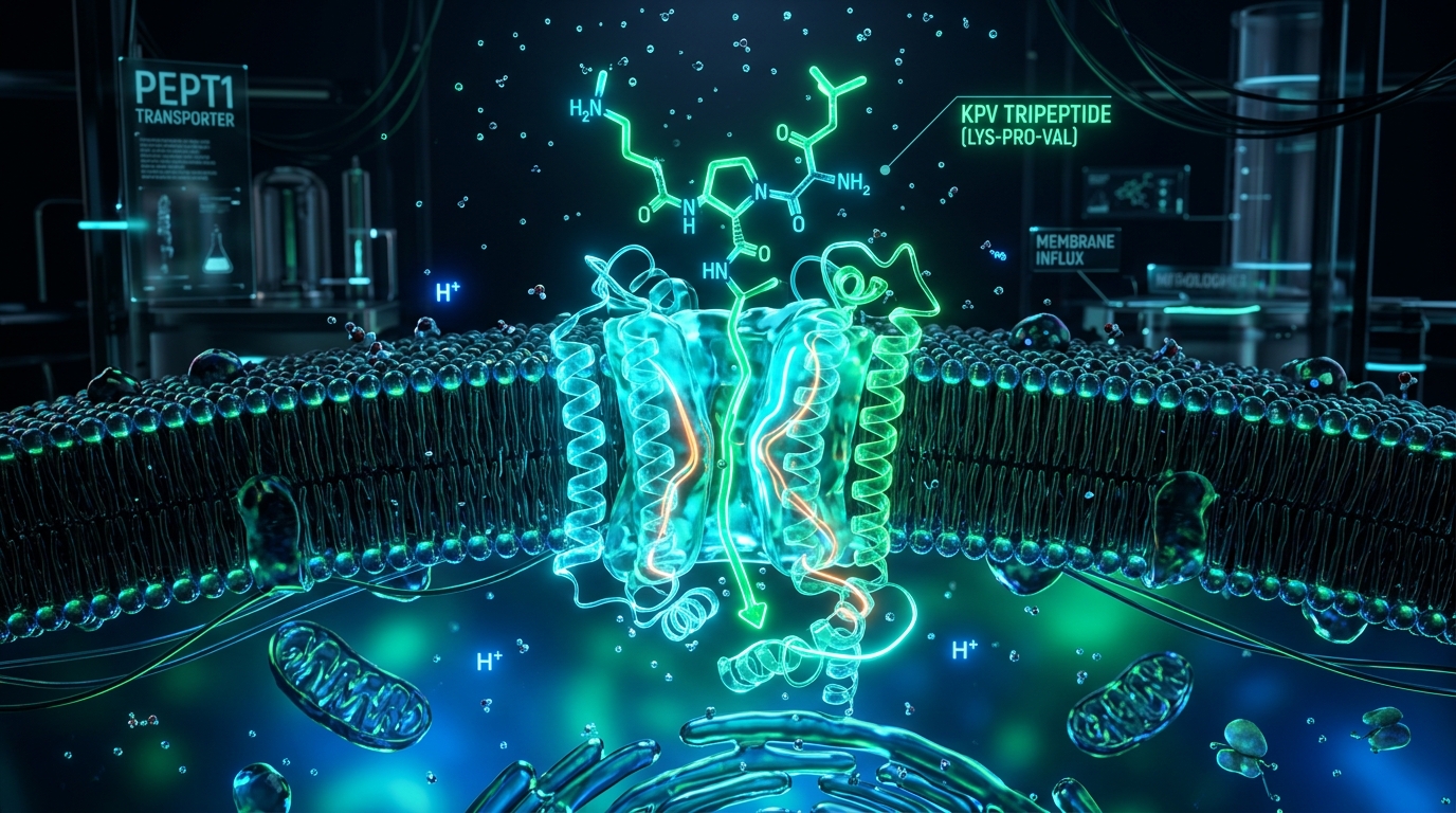
The most concrete finding comes from Dalmasso and colleagues, published in Gastroenterology in 2008. PepT1 (SLC15A1) is a proton-coupled di- and tripeptide transporter normally expressed in the small intestine and induced in colonic epithelium during intestinal inflammation. Working in human intestinal epithelial lines Caco2-BBE and HT29-Cl.19A and in Jurkat T cells, the group used unlabelled KPV as a competitor against a radiolabelled PepT1 substrate, and separately used tritiated KPV to establish uptake kinetics. Both approaches supported PepT1-mediated transport of KPV into epithelial and immune cells.
This separates KPV from the receptor-agonist model. The proposal is not that KPV binds a surface receptor and triggers a cascade, but that a transporter carries it into the cytosol where it interferes with signalling directly.
The transporter's inflammation-associated expression pattern is worth noting carefully. Because PepT1 is induced in colonic epithelium during inflammation, the transport findings may provide a mechanistic basis for investigating whether KPV uptake differs under inflammatory conditions. That is a research hypothesis and mechanistic implication raised by existing data, not a demonstrated tissue-targeting property in an intact organism.
NF-κB and MAPK Signalling
In the same work, nanomolar KPV inhibited NF-κB activation in cytokine-stimulated cells. The readouts were an NF-κB luciferase reporter construct, Western blots tracking IκB-α degradation and phosphorylation over time, quantitative RT-PCR and ELISA. In Caco2-BBE cells stimulated with IL-1β, 10 nM KPV reduced the inflammatory response across those measures. The same concentrations inhibited MAP kinase signalling alongside NF-κB, though the MAPK finding is consistently less developed in the literature and is better read as a co-observation than as an independently characterized pathway. No study located isolates a specific MAPK branch as a primary route of KPV activity.
FDA's reviewers describe NF-κB inhibition, leading to reduced proinflammatory cytokine expression, as one of two proposed contributors to KPV's anti-inflammatory properties. The second, drawing on Getting and on Mandrika, is direct inhibition of the effects of proinflammatory cytokines such as IL-1β, independent of any effect on their expression. Whether these are two descriptions of one mechanism or two separate activities has not been resolved.
Melanocortin Receptor Independence
This is where the evidence is unusually clean, and where a large share of commercial KPV content gets it backwards. Available evidence argues against melanocortin-receptor-mediated activity as the primary explanation for KPV's observed effects.
FDA's assessment assembles four independent lines. KPV failed to displace radiolabelled alpha-MSH binding in rat brain tissue, murine melanoma cells and MC1R-expressing murine macrophages, across work by Lyson (1994), Mandrika (2001) and Tatro and Entwistle (1994). Unlike alpha-MSH, KPV did not raise cyclic AMP in MC1R-expressing murine macrophages. And pharmacological and genetic approaches together failed to demonstrate MC2, MC3 or MC4 involvement in KPV's anti-inflammatory or wound-healing effects.
The animal work agrees. Kannengiesser and colleagues found that in MC1R-deficient (MC1Re/e) mice, KPV treatment rescued all treated animals from death during DSS colitis, which is difficult to reconcile with an MC1R-dependent mechanism. Their conclusion was that the effects appear at least partially independent of MC1R signalling.
Any page claiming KPV works through MC1R and MC3R activation is stating something the primary literature specifically tested and did not support.
KPV in Gut & Mucosal Research
Gut and mucosal work is where KPV peptide research is deepest, and where the gap between what was studied and what gets claimed is widest.
Intestinal Epithelial Models
The cellular work centres on two human intestinal epithelial lines, Caco2-BBE and HT29-Cl.19A, with Jurkat cells as an immune-compartment comparator. These are transformed lines grown in culture. They model epithelial signalling behaviour usefully. They do not model an intact intestine, a microbiome, or an immune system.
Animal Colitis Models
Dalmasso's group extended the cellular findings into two induced colitis models in mice. KPV was delivered in drinking water, and inflammation was assessed histologically and by proinflammatory cytokine mRNA expression. Orally delivered KPV reduced the incidence of both DSS-induced and TNBS-induced colitis, with the reduction reflected in cytokine expression.
Kannengiesser and colleagues, publishing in Inflammatory Bowel Diseases the same year, used a different pairing: DSS colitis and CD45RBhi transfer colitis. In the DSS model, KPV-treated mice recovered earlier and regained significantly more body weight. Histological inflammatory infiltrates were significantly reduced, corroborated by significantly lower myeloperoxidase activity in colonic tissue. In the transfer colitis model, KPV treatment produced recovery, weight regain and reduced histological inflammatory change.
DSS and TNBS are chemically induced colitis models. They are standard tools and they are informative, but they are not inflammatory bowel disease. Transfer colitis introduces a T-cell-driven component and sits closer to chronic immune-mediated colitis, which is why the two-model design in each study carries more weight than either alone. Consistency of direction across three models, two laboratories and three cell lines is the strongest feature of the KPV literature.
Reading Delivery-System Results Correctly
A substantial share of KPV work published after 2008 concerns engineered delivery: the peptide loaded into nanoparticles, hydrogels or functionalised carriers designed to survive transit and concentrate it at inflamed tissue. This exists because free KPV is a small, unprotected peptide subject to rapid peptidase degradation. Results from that work are frequently quoted as though they describe KPV itself, but an outcome produced by a peptide inside a targeted carrier is a property of the delivery system as much as of the peptide. When evaluating any KPV performance claim, the first question is what was actually administered.
KPV Research Evidence: Human, Animal and In Vitro
The table below separates the KPV literature by evidence level, because the most common error in writing about this compound is letting a cell-culture result read like a clinical one. Each row states the model used, what was measured, and what the result does not extend to.
| Evidence level | Main model | Research question | Current finding | Major limitation |
|---|---|---|---|---|
| Human clinical | None identified | Efficacy or safety in humans | No published studies of KPV administered to humans by any route | The category is empty |
| Human PK/PD | None identified | Absorption, distribution, clearance | No pharmacokinetic or toxicokinetic studies identified by FDA | No basis for translating preclinical concentrations to human exposure |
| Ex vivo human tissue | Cadaver skin | Does KPV cross skin? | Poor permeation; iontophoresis and microneedle abrasion increased penetration below the stratum corneum | In vitro cadaver skin. Silent on systemic absorption in vivo |
| Animal | DSS and TNBS colitis, mice | Does orally delivered KPV reduce colitis? | Reduced incidence of both, with reduced proinflammatory cytokine expression | Chemically induced models. Not IBD |
| Animal | DSS and CD45RBhi transfer colitis, mice | Anti-inflammatory potential and mode of action | Earlier recovery, greater weight regain, reduced infiltrates and MPO activity; MC1R-deficient treated animals survived | Two induced models; MC1R independence described as partial |
| Animal | Rabbit corneal epithelium | Effect on epithelial wound healing | Effects on corneal epithelial wound closure reported, with a nitric oxide role | Single tissue, single species |
| In vitro | Caco2-BBE, HT29-Cl.19A, Jurkat | Uptake route and signalling effect | PepT1-mediated uptake; nanomolar KPV inhibited NF-κB and MAPK activation | Transformed cell lines. No barrier, microbiome or immune context |
| In vitro, receptor | Rat brain tissue, murine melanoma cells, MC1R-expressing macrophages | Does KPV act at melanocortin receptors? | No displacement of radiolabelled α-MSH; no cAMP increase; MC2/MC3/MC4 involvement not demonstrated | Negative findings, which constrain mechanism rather than explain it |
| Nonclinical safety | None identified | Acute, repeat-dose, genotoxicity, DART, carcinogenicity | FDA identified no studies in any category | The nonclinical toxicology package is absent |
Read top to bottom, the pattern is clear: the mechanistic and animal tiers are populated and mutually corroborating, while the human and nonclinical-safety tiers are empty. That shape, more than any single finding, defines the current state of KPV peptide research.
KPV vs BPC-157 vs Thymosin Alpha-1
These three are frequently grouped in catalogs and clinic marketing. From a research standpoint they are not comparable objects, and the differences determine what a given sourcing decision commits a laboratory to. Laboratories evaluating mucosal barrier and immune signaling research bundles frequently reference our verified Gut & Mucosal Panel to study these three compounds alongside one another.
| Feature | KPV | BPC-157 | Thymosin alpha-1 |
|---|---|---|---|
| Molecular context | Tripeptide, 342.43 g/mol free base; C-terminal fragment of alpha-MSH | Pentadecapeptide; sequence derived from a gastric juice protein | 28-residue N-terminally acetylated peptide; fragment of prothymosin alpha |
| Reported mechanisms | PepT1-mediated uptake; NF-κB and MAPK inhibition; molecular target unknown | Multiple proposed pathways; no consensus target | Signalling via Toll-like receptors on dendritic cells and macrophages; T-cell maturation effects |
| Typical research questions | Mucosal and intestinal inflammatory signalling; epithelial wound models | Tissue repair, angiogenesis, gastrointestinal mucosal injury | Immune modulation, T-cell function, adjuvant settings |
| Highest evidence level | Rodent colitis and wound models; human cell lines | Rodent models across several tissue types | Human randomized trials; marketed as thymalfasin (Zadaxin) in 35+ countries for chronic hepatitis B |
| Human data | Human translational evidence has not been established | None establishing efficacy for the use FDA evaluated | Substantial, including published human pharmacokinetics |
| US regulatory status | Not FDA-approved; not on 503A Bulks List | Not FDA-approved; not on 503A Bulks List | Not FDA-approved in the US; has held FDA orphan designations |
| July 2026 PCAC | Reviewed for wound healing and inflammatory conditions | Reviewed for ulcerative colitis | Not among the seven substances reviewed |
The distinction that matters is evidentiary rather than comparative. Thymosin alpha-1 has been administered to humans in controlled studies and human pharmacokinetic parameters exist in the published record; KPV and BPC-157 have not and do not. Grouping all three as "research peptides" is accurate as a supply-chain description and misleading as a scientific one. None of this indicates which compound anyone should use.
Is KPV FDA Approved? The 2026 Regulatory Update
No. KPV is not an FDA-approved drug. KPV is not currently on the FDA's Section 503A Bulks List, and the July 2026 PCAC recommendation did not itself establish eligibility for compounding under Section 503A.
That answer needs the chronology behind it, because the July 2026 meeting has been widely misreported.

How the 503A Pathway Works
Under section 503A of the Federal Food, Drug, and Cosmetic Act, a licensed pharmacist or physician may compound a drug for an individual patient using a bulk drug substance only if that substance is a component of an FDA-approved drug, has a USP or NF monograph, or appears on the 503A Bulks List. KPV meets none of the three conditions.
The Nomination and FDA's Review
KPV was nominated for the 503A Bulks List by Wells Pharmacy Network (docket document FDA-2015-N-3534-0294). The nomination was later withdrawn, and FDA elected to evaluate the substances on its own initiative. The package was internally inconsistent about whether it covered free base or acetate, and FDA noted that the certificate of analysis submitted with it referred to one substance by name in the title and a different one by molecular formula. FDA treated both forms as distinct substances and evaluated each separately.
The evaluated uses were wound healing and inflammatory conditions, in a proposed 0.1% topical cream or gel. Not injection, not oral, not nasal spray.
FDA staff, in an assessment dated May 12, 2026, proposed not adding either form to the list. Their reasoning had four parts: both forms were deemed not well-characterized, on grounds of inconsistent naming conventions and missing quality-control attributes including impurity profile, aggregation and microbiological testing; the extent of historical compounding use was unknown, with outsourcing facilities reporting no KPV products from January 2017 through June 2025; there was no evidence of effectiveness, since human translational evidence has not been established; and there was no clinical or nonclinical safety information, with immunogenicity and aggregation risk specifically unassessed. FDA also weighed the existence of approved therapies for wound healing and for inflammatory conditions such as psoriasis and eczema.
What Happened at the July 2026 Meeting
The Pharmacy Compounding Advisory Committee met July 23-24, 2026 at FDA's White Oak campus under docket FDA-2025-N-6895. KPV free base and KPV acetate were voted on separately during the July 23 morning session, alongside BPC-157. Across the two days the committee reviewed seven substances and, on split votes, recommended six of them for the 503A Bulks List. Emideltide (DSIP) was the only one not recommended. KPV was among the six.
The PCAC recommendation directly contradicted the agency's own staff assessment, and FDA reviewers restated their concerns at the meeting.
What the Recommendation Did and Did Not Change
Advisory committee recommendations are non-binding. FDA generally follows them but is not legally required to.
Legally, nothing changed. Holland & Knight's August 2026 analysis states the position plainly: the PCAC recommendation did not result in any change in law, these peptides still cannot be lawfully compounded, and FDA can continue to take enforcement action against compounding pharmacies that make them. The only routes to a different outcome are formal notice-and-comment rulemaking or an act of Congress amending the FDCA.
The accurate framing is therefore: PCAC recommendation ≠ FDA approval ≠ final agency action ≠ 503A eligibility. A favourable vote is a procedural step in a process that has not concluded.
For the broader 2026 regulatory picture across peptide sourcing generally, including the objective intent doctrine and RUO supplier obligations, see our 2026 peptide sourcing and FDA regulatory guide.
What Should Laboratories Verify Before Sourcing KPV?
FDA's characterization finding is the most useful sourcing document a KPV buyer currently has, because it describes precisely what commercial documentation for this compound has typically failed to show.
Identity, and which identity. Establish at the quotation stage whether you are buying free base or acetate. They differ in molecular weight (342.43 versus 402.5 g/mol), in water solubility, and in what a mass-spectrometric identity check should return. Because the same CAS number is commonly applied to both in public references, CAS alone will not resolve this. Ask for the molecular formula and stated salt form in writing.
Lot number and batch traceability. A certificate that cannot be tied to the specific vials in your shipment is a marketing document. The lot on the paperwork must match the lot on the container.
What the Analytical Methods Can and Cannot Show
HPLC. Reversed-phase HPLC separates and quantifies the main peak against detected impurities under a given method, which supports a purity figure. It does not confirm identity on its own, and it will not detect species that do not absorb at the monitored wavelength or do not elute under the method used.
LC-MS. LC-MS confirms molecular mass and supports identity. It is the check that would catch a free base and acetate mix-up, or a truncated sequence. It does not quantify purity, and mass alone will not distinguish stereoisomers.
Neither method. Neither addresses biological activity, and neither addresses peptide aggregation, which FDA flagged specifically and which may require size exclusion chromatography or field flow fractionation. Microbiological quality and bacterial endotoxin are separate assays again. Endotoxin is not visible to HPLC or LC-MS and matters for cell-based work, particularly immunological readouts, where contamination can generate exactly the inflammatory signal an experiment is measuring.
Degradation behaviour. Under forced degradation including acid and alkaline hydrolysis and oxidation with hydrogen peroxide, KPV acetate has been reported to form lysine-proline-diketopiperazine as a major degradation product, with rapid breakdown to the diketopiperazine plus free proline and valine under peroxide. Useful context when specifying storage and interpreting an unexpected peak.
Consistency, Origin and Documentation
For any study running longer than a single lot, the impurity profile needs to stay stable across reorders, not just the headline purity number. FDA's briefing document notes that solid-phase peptide synthesis generates characteristic impurities from incomplete coupling, truncation and side reactions, plus residual solvents, coupling reagents and scavengers. A supplier that changes synthesis route between lots may hold the same purity figure while shifting the impurity profile beneath it. Ask whether the route and the testing laboratory are fixed.
An institutional procurement file should capture the specification required including salt form, the analytical evidence required per lot, who reviews incoming certificates and against what acceptance criteria, the retention policy for certificates and lot records, and the RUO handling declaration.
On purity thresholds. 99 Purity Wholesale specifies ≥99.0% purity for KPV, verified by HPLC and LC-MS through Freedom Diagnostic, with a batch-matched certificate of analysis per order. That is a supplier specification: not a legal requirement, not a regulatory standard for research materials, and not evidence of biological activity. Document the specification your own application requires rather than assuming a market standard exists.
How to Evaluate a KPV COA
FDA's review of publicly available KPV documentation produced a finding worth restating: most certificates located for KPV free base contained only a purity result, with no impurity limits or results demonstrating control of the impurity profile.
A certificate that supports a procurement decision should let you check:
- Lot identification, matching the material in hand
- Product identity, stated as free base or acetate, with molecular formula
- Test date and testing laboratory, named and independent of the manufacturer
- HPLC purity result, with the method stated; a chromatogram is more informative than a number
- LC-MS identity evidence, confirming molecular mass against the stated form
- Related substances, meaning individual and total impurity limits with actual results, not an assay figure alone
- Water content and counter-ion content where an acetate is supplied
- Additional testing performed, such as microbiological or endotoxin results
The limitations matter as much as the contents. A COA reports what was tested, on the sample tested, by the method used. It is not a guarantee of stability in your hands, it does not establish biological activity, and where aggregation and microbiological attributes are untested it is silent on them rather than reassuring.
Our guide to reading a peptide certificate of analysis covers the general methodology, and the supplier verification guide covers the diligence process around it.
What KPV Research Still Does Not Tell Us
Three absences deserve naming beyond what the evidence table records, because each one carries a decision consequence.
No human exposure data of any kind. FDA searched PubMed, Embase, ClinicalTrials.gov, DailyMed and Drugs@FDA and found no studies of KPV administered to humans by any route, and no pharmacokinetic or toxicokinetic studies for either form. Human translational evidence has not been established. There is therefore no basis for relating a concentration effective in a Caco2-BBE monolayer to any human exposure. The absence of adverse event reports in FDA's surveillance databases reflects the absence of documented human use, not a demonstration of safety.
Immunogenicity is unassessed. FDA flagged it specifically: peptides can provoke immune responses, including neutralizing antibody responses against endogenous counterparts, and aggregation is a recognized risk factor. This is the gap most relevant to anyone extrapolating from rodent work.
The molecular target is unidentified. PepT1 uptake is supported and melanocortin receptor involvement is largely excluded, which narrows the field considerably, but the target through which KPV inhibits NF-κB remains unknown.
KPV Procurement for Research Laboratories
99 Purity Wholesale supplies KPV under Cellular Health Research as a lyophilized solid powder for research use only, with independent HPLC and LC-MS verification through Freedom Diagnostic and a batch-matched certificate of analysis. For protocols requiring liquid formulations, laboratories can also evaluate pre-dissolved options on the KPV Spray product page. Specifications, kit sizes and minimum order quantity for standard vials are on the primary KPV product page; wholesale accounts require a business EIN and a research-use declaration via the wholesale application.
Institutional buyers should document their RUO position explicitly. KPV is not an approved drug and, following the July 2026 PCAC meeting, KPV is not currently on the FDA's Section 503A Bulks List. Material purchased as a research reagent should be handled, stored and recorded as one.
The KPV literature illustrates why that discipline pays off. The science is interesting, the preclinical signal is consistent across two independent groups, and the conclusion is still that human translational evidence has not been established. A supply relationship producing documentation you can actually inspect is the part of that picture a laboratory controls.
