
What's the Difference Between Fluorescence, Chemiluminescence, and Bioluminescence?
In analytical biochemistry, molecular biology, and cellular assay development across our wholesale research compounds catalog, light-emitting phenomena serve as primary detection modalities. While fluorescence, chemiluminescence, and bioluminescence all yield optical signals measurable by photodetectors, their underlying physical, chemical, and biological mechanisms differ fundamentally.
To establish clear terminology in our scientific glossary, all three techniques fall under the broad umbrella of luminescence—the emission of light from a substance not resulting from heat (often termed "cold light," distinguishing it from incandescence). Within luminescence:
- Photoluminescence encompasses light emission triggered by the absorption of optical photons, divided into fluorescence (rapid radiative emission) and phosphorescence (delayed spin-forbidden emission).
- Chemiluminescence describes light produced directly by an exergonic chemical reaction without photon input.
- Bioluminescence is a specialized biological form of chemiluminescence where an enzyme catalyzes the light-emitting chemical reaction within a living organism or biological preparation.
Understanding where excitation energy originates—from external photon absorption, chemical bond rearrangement, or enzymatic catalysis—explains the operational differences researchers encounter in instrumentation design, background signal, kinetic behavior, and sample compatibility under strict laboratory compliance standards.
What Is Fluorescence?
Fluorescence occurs when a molecule (a fluorophore) absorbs a photon of light at a specific excitation wavelength, promoting an electron from its singlet ground state (S₀) to an excited electronic state (S₁ or higher). Within picoseconds to nanoseconds, the molecule undergoes internal conversion and vibrational relaxation to the lowest vibrational level of S₁. From there, the molecule relaxes back to the S₀ ground state by emitting a photon of lower energy—and consequently longer wavelength—than the absorbed photon.
The difference in energy and wavelength between the absorption maximum and the emission maximum is known as the Stokes shift. This wavelength displacement allows optical instruments to isolate the emitted fluorescent signal from the excitation light path using spectral filters or monochromators.
[Ground State S₀] + Photon (Excitation) ──> [Excited State S₁]
                                                    │
                                           Vibrational Relaxation
                                                    │
                                                    ▼
[Ground State S₀] <── Photon (Emission) ── [Relaxed S₁ State]
Key Technical Characteristics of Fluorescence
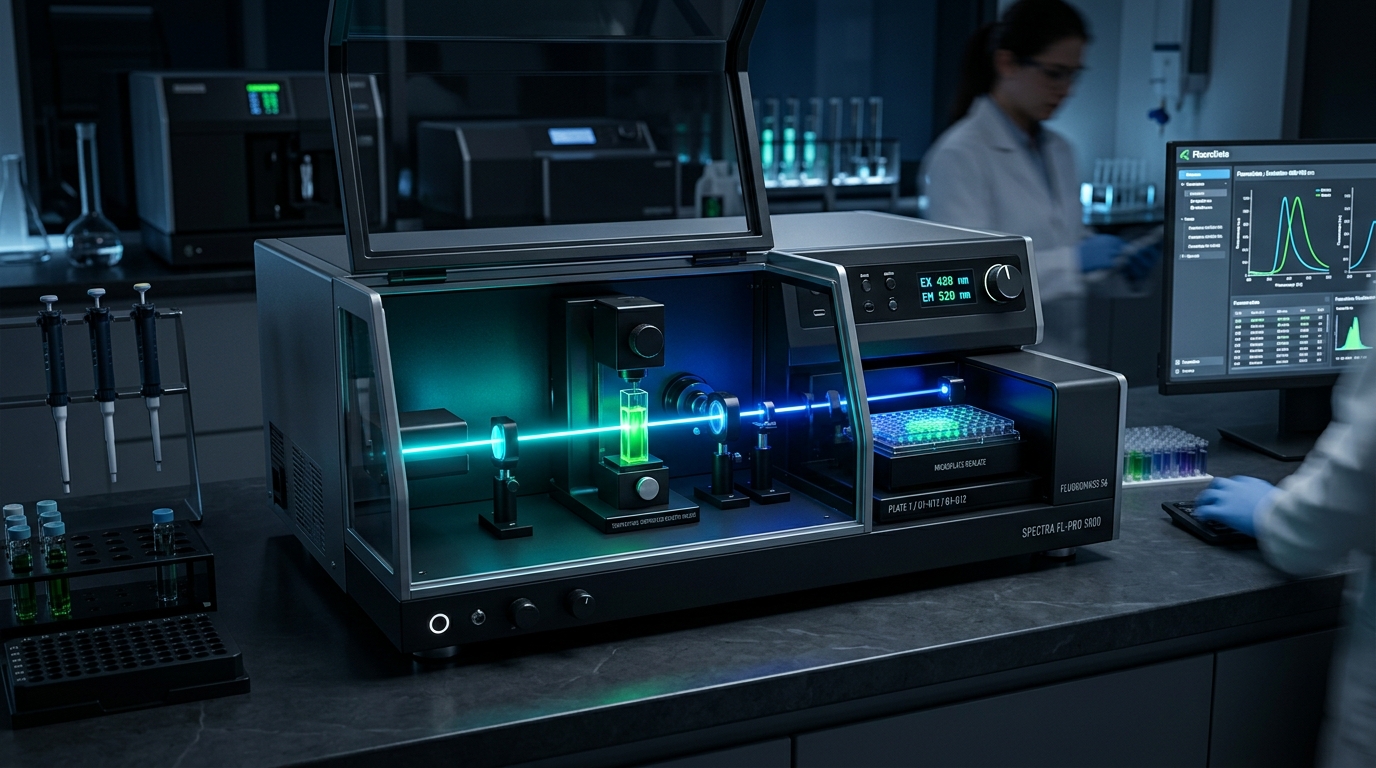
- Requirement for External Illumination: Fluorescence fundamentally depends on a continuous or pulsed external light source (such as a xenon arc lamp, light-emitting diode [LED], or laser).
- Nanosecond-Scale Emission Kinetics: The fluorescence lifetime of typical organic fluorophores and fluorescent proteins ranges from 1 to 10 nanoseconds. Consequently, emission is effectively instantaneous upon illumination and persists continuously as long as excitation light is maintained.
- Spectral Flexibility: A vast library of fluorophores exists—spanning ultraviolet, visible, and near-infrared (NIR) spectrums—including small molecule dyes (FITC, Alexa Fluors, Cyanine dyes), fluorescent proteins (GFP, mCherry), and quantum dots. Multi-color multiplexing is straightforward when fluorophores have distinct excitation and emission profiles.
Background and Operational Considerations
Because fluorescence requires illuminating the sample with excitation light, optical background signal can present a major analytical challenge. Excitation light can scatter off sample components (Rayleigh and Raman scattering) or excite endogenous molecules present in biological matrices—such as flavins, NADH, collagen, cell culture media, and plastic microplates—generating autofluorescence.
In clean in vitro solutions or isolated cell monolayers, autofluorescence is manageable through proper optical filtering and baseline subtraction. When working with reconstituted peptides, verifying baseline purity with independent Certificates of Analysis (COAs) and following a proper reconstitution and storage protocol guide ensures background interference is kept to a minimum.
What Is Chemiluminescence?
Chemiluminescence is the emission of light resulting from a chemical reaction. Instead of using external photon absorption to elevate electrons to an excited state, an exergonic chemical reaction—typically involving oxidation—directly generates a reaction intermediate or product in an electronically excited state. As this excited species relaxes to its ground state, it releases the excess energy as visible or near-visible light.
A classic laboratory example is the oxidation of luminol in the presence of an oxidant (such as hydrogen peroxide) and a catalyst (such as horseradish peroxidase [HRP] or iron complexes). In Enhanced Chemiluminescence (ECL) reagents used for protein detection, chemical enhancers (like substituted phenols or boronic acids) are added to increase light output intensity and extend signal duration, as explored in our COA verification guide.
Key Technical Characteristics of Chemiluminescence
- Zero External Optical Excitation: Chemiluminescence requires no light source, excitation filters, or illumination optics. The light generated by the sample originates entirely from chemical potential energy.
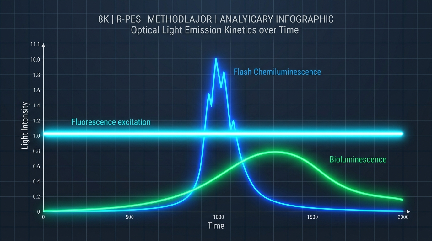
- Reaction-Driven Signal Kinetics: Unlike the steady-state emission of fluorescence under continuous illumination, chemiluminescent signals exhibit time-dependent kinetic profiles. Flash chemiluminescence produces a rapid spike of light lasting seconds upon reagent injection, while glow chemiluminescence maintains a sustained light output over minutes to hours as the substrate is consumed.
- Low Excitation-Related Background: By eliminating external excitation light, chemiluminescence avoids Rayleigh/Raman scattering and excitation-induced matrix autofluorescence. The photodetector records only light generated by the chemical reaction itself.
Background and Operational Considerations
While chemiluminescence eliminates optical autofluorescence, it introduces chemical background considerations. Non-specific chemical oxidation, thermal decomposition of reagents, or trace metallic impurities in buffers can cause background light emission (autoxidation). Evaluating reagent purity according to rigorous bulk supplier evaluation criteria ensures reproducible quantitative measurements.
What Is Bioluminescence?
Bioluminescence is light production carried out by biological systems, mediated by enzymatic catalysis. Mechanistically, bioluminescence is an enzyme-catalyzed subcategory of chemiluminescence occurring naturally in diverse organisms, including fireflies, marine dinoflagellates, jellyfish, copepods, and certain bacteria and fungi.
In standard bioluminescent reactions, an enzyme broadly termed a luciferase catalyzes the oxidation of a small-molecule substrate termed a luciferin. Depending on the evolutionary origin of the system, the reaction may require cofactors such as adenosine triphosphate (ATP), magnesium ions (Mg²⁺), or molecular oxygen (O₂).
For example, the firefly luciferase (Photinus pyralis) reaction proceeds as follows:
Luciferin + ATP + O₂  ──(Luciferase, Mg²⁺)──>  Oxyluciferin* + AMP + PPi + CO₂

Oxyluciferin*  ──────────────>  Oxyluciferin + Photon (Light, λmax ≈ 560 nm)
Other systems, such as Renilla (sea pansy) or Gaussia luciferases, utilize coelenterazine as a substrate and do not require ATP, enabling ATP-independent reporter assays often used alongside NAD+ longevity research studies.
Key Technical Characteristics of Bioluminescence
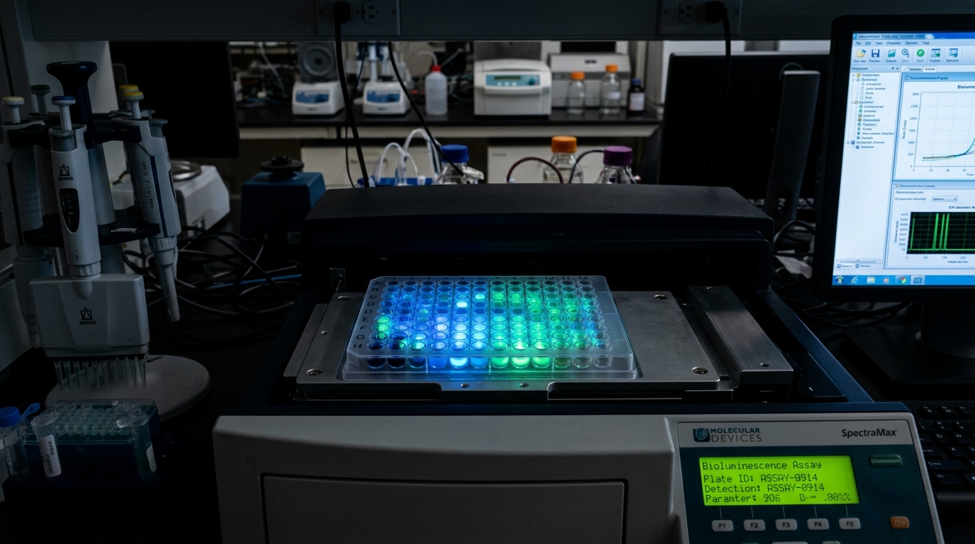
- Enzymatic Specificity & Biological Origin: Bioluminescent reactions rely on highly specific enzyme-substrate pairings. Mammalian cells and tissues do not endogenously express luciferases or synthesize luciferin substrates, providing an essentially zero biological background baseline.
- Genetic Encoding: In molecular research, luciferase genes can be cloned into expression vectors under the control of specific promoters, enhancers, or response elements. This allows bioluminescence to serve as a direct reporter for transcriptional activity, gene regulation, signal transduction, or cell survival, such as evaluating receptor activation in Semaglutide vs. Tirzepatide comparative research.
- Substrate Dependence & Kinetics: Light emission requires supplying the exogenous luciferin substrate. Depending on the luciferase system and formulation (flash vs. steady-glow buffers), light output can range from rapid transient bursts to stable glow signals lasting several hours.
Is Bioluminescence a Form of Chemiluminescence?
Mechanistically and chemically, yes: bioluminescence is an enzyme-catalyzed variant of chemiluminescence. Both phenomena generate electronically excited states through exergonic chemical reactions without requiring photon absorption.
However, in practical laboratory terminology, researchers consistently treat them as distinct detection toolkits:
| Practical Metric | Chemiluminescence (Non-Enzymatic / Synthetic) | Bioluminescence (Enzyme-Mediated) |
|---|---|---|
| Primary Reagents | Synthetic substrates, oxidizing agents, chemical catalysts (e.g., Luminol, Acridinium, HRP) | Biological enzymes (Luciferase) and organic substrates (Luciferin, Coelenterazine) |
| Genetic Applicability | Cannot be genetically encoded; added as chemical detection reagents | Luciferase enzymes can be genetically encoded into host genomes or plasmids |
| Typical Assay Format | End-stage detection reagents added to cell lysates, membranes, or microplates | Live-cell assays, cell-free viability assays, promoter reporters, and live-animal imaging |
Defining bioluminescence as a specialized biological class of chemiluminescence preserves chemical accuracy while respecting practical laboratory distinctions.
Fluorescence vs. Chemiluminescence vs. Bioluminescence Comparison
The following table summarizes the core physical, chemical, and operational differences across all three analytical modalities:
| Parameter | Fluorescence | Chemiluminescence | Bioluminescence |
|---|---|---|---|
| Light Source Mechanism | Photoluminescence: absorption of excitation photon elevates fluorophore to excited singlet state | Chemiluminescence: exergonic chemical reaction yields product in excited electronic state | Biological Chemiluminescence: enzyme (luciferase) catalyzes oxidation of substrate (luciferin) to excited state |
| External Excitation Light Needed? | Yes (xenon lamp, LED, or laser required) | No (purely chemical light production) | No (purely enzymatic light production) |
| Biological Requirement | No biological components required; compatible with synthetic dyes, proteins, and inorganic tags | Purely chemical reaction; may utilize coupled enzymes (e.g., HRP) as catalysts | Requires functional enzyme-substrate reaction pair; compatible with cell-free or intact biological systems |
| Temporal & Kinetic Profile | Continuous, steady-state signal maintained while illumination is active; instantaneous decay | Reaction-dependent kinetics; flash (seconds) or sustained glow (minutes to hours) as substrate converts | Reagents/substrate-dependent kinetics; flash or glow profile determined by enzyme turnover and substrate availability |
| Background Signal Constraints | Excitation light scattering and matrix autofluorescence can limit signal-to-noise in complex samples | No optical autofluorescence; background limited by chemical autoxidation or non-specific reagent breakdown | Extremely low biological background in mammalian systems lacking endogenous luciferase activity |
| Typical Instrumentation | Spectrofluorometer, fluorometer, fluorescence microplate reader, flow cytometer, confocal microscope | Luminometer, chemiluminescent plate reader, CCD gel/membrane imaging system | Luminometer, bioluminescent plate reader, highly sensitive cooled CCD in vivo imaging system (IVIS) |
| Common Research Applications | Flow cytometry, live-cell imaging, qPCR, fluorescent immunoassays, protein/DNA quantification | Western blot ECL detection, chemiluminescent immunoassays (CLIA), reactive oxygen species (ROS) assays | Promoter reporter assays, ATP cell viability assays, protein interaction (BRET), non-invasive in vivo animal imaging |
| Primary Operational Limitation | Autofluorescence in tissue/media; photobleaching of fluorophores under intense illumination | Signal decays as reagents consume; restricted wavelength tuning compared to fluorophore selection | Requires substrate delivery; quantitative signal depends on substrate diffusion, ATP, and oxygen availability |
How Do the Three Methods Differ in Practice?
Beyond basic definitions, several practical operational factors dictate how these methods perform in real-world research workflows.
1. Background Signal and Signal-to-Noise Behavior
In optical detection, signal-to-noise ratio (S/N) is often more important than absolute signal intensity.
- Fluorescence: Because excitation light passes directly into or through the sample matrix, any light scattering or endogenous fluorophores (such as cellular flavins, aromatic amino acids, or cell culture media components like phenol red) will produce background light. Filtering and spectral unmixing reduce this noise, but autofluorescence remains a primary consideration in whole-blood, tissue, or thick specimen imaging.
- Chemiluminescence & Bioluminescence: By omitting excitation illumination, these modalities eliminate excitation-driven autofluorescence entirely. The detector observes a dark background interrupted only by light produced from the reaction itself. As a result, luminescence methods frequently achieve superior sensitivity for low-abundance targets, even if absolute photon output is lower than a strongly illuminated fluorescent dye.
2. Temporal Behavior and Measurement Kinetics
The timing of signal acquisition varies significantly across modalities:
- Fluorescence supports continuous, real-time monitoring over arbitrary timeframes. As long as excitation light is applied and photobleaching is minimized, signal intensity can be recorded continuously or sampled repeatedly at high frequencies. When measuring molar concentration and dilution factors, using our interactive peptide dosage & reconstitution calculator simplifies experimental design.
- Chemiluminescence and Bioluminescence are dynamic chemical reactions. Light output rises as reagents mix, reaches a peak, and decays as substrate is depleted or enzyme undergoes suicide inactivation. Assays must be calibrated around specific read windows (e.g., reading 10 minutes post-substrate addition) or integrated over defined time intervals.
3. Multiplexing Capabilities
- Fluorescence excels at multi-target multiplexing. Modern flow cytometers and fluorescence microscopes routinely resolve 10 to 30 distinct color channels simultaneously by pairing narrow-band excitation sources with specific emission filters.
- Luminescence multiplexing is more constrained because emission spectra of chemiluminescent substrates and luciferases are relatively broad. However, dual-luciferase assays (e.g., pairing Firefly luciferase with Renilla or Nanoluc luciferase utilizing distinct substrates and emission wavelengths) allow robust two-target normalization in a single sample well.
What Equipment Is Used for Fluorescence Detection?
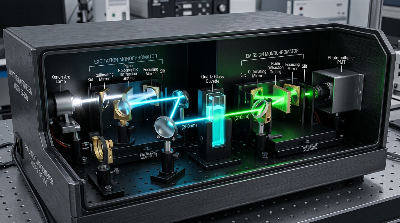
Analytical terminology for fluorescence equipment can be confusing because manufacturers and literature frequently use overlapping terms. Understanding what an instrument actually does requires looking at its optical components.
Clarifying Instrument Terminology
- Fluorescence Spectrometer / Fluorescence Spectrophotometer: These terms are functionally synonymous in modern laboratory usage. A fluorescence spectrophotometer features both an excitation monochromator and an emission monochromator. This permits continuous scanning across a range of wavelengths to generate full excitation spectra (varying excitation while measuring fixed emission) and emission spectra (fixing excitation while scanning emission).
- Spectrofluorometer: A term used interchangeably with fluorescence spectrophotometer in peer-reviewed literature. It explicitly denotes an instrument designed for high-resolution spectral scanning rather than single-wavelength filter measurements.
- Fluorimeter / Fluorometer: Historically and in current product naming, "fluorometer" often refers to simpler, filter-based detection devices (such as compact benchtop quantitation units like the Qubit). Filter fluorometers use fixed optical filters rather than scanning monochromators. They offer high sensitivity and lower cost for dedicated single-wavelength assays, but lack spectral scanning flexibility.
How Do Researchers Choose Between Fluorescence, Chemiluminescence, and Bioluminescence?
Selecting the optimal detection modality requires matching experimental requirements with the physical strengths of each technique, particularly for analytical labs and medical spas following compounding pharmacy sourcing guidelines:

- Is matrix autofluorescence a limiting factor? If working with intact tissues, blood, or whole animal imaging where excitation light causes severe background interference, bioluminescence or chemiluminescence is preferred due to superior signal-to-noise ratios.
- Is real-time continuous tracking required? If an experiment demands continuous, multi-hour tracking of a target without adding external substrates at defined intervals, fluorescence provides a steady-state signal under illumination.
- Is genetic modification feasible? If host cells can be stably or transiently transfected, genetically encoded luciferase reporter systems offer highly quantitative, low-background readouts for signaling pathways. If genetic engineering is impossible or undesirable, synthetic fluorescent labels or chemiluminescent reagents are used.
- What instrumentation is available? While multi-mode microplate readers often support fluorescence, chemiluminescence, and bioluminescence in a single chassis, dedicated research facilities applying for a wholesale B2B application can equip their laboratories with dedicated optical hardware suited to their volume.
